
Practical Management of Atopic Dermatitis: Nurse Practitioner and Physician Assistant Perspectives - Episode 2
Differences in Atopic Dermatitis Clinical Presentation
The differences in early skin immune changes in pediatric atopic dermatitis (AD) versus long-standing adult AD and variations in the condition’s presentation among patients of color.
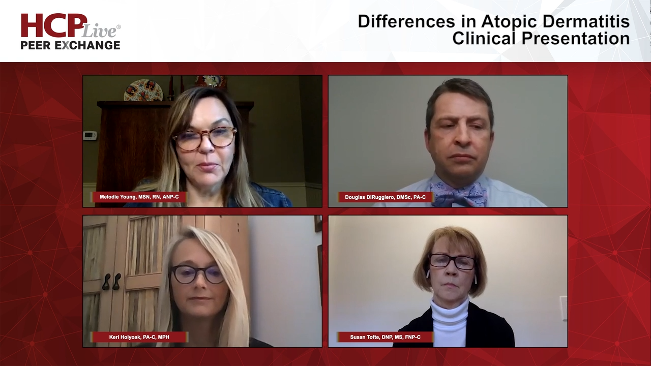
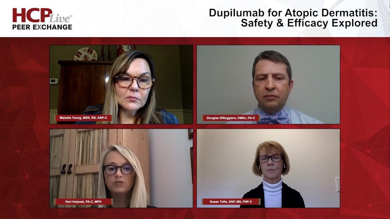
Melodie Young, MSN, RN, ANP-C: How do you communicate this information to your patients? Sometimes I’ll say, “Your skin’s job is to keep the good stuff in and the bad stuff out. When you have this disease, that’s not what is happening.” Keri or Susan, could you speak to how the atopic dermatitis presents in a dermatology clinic?
Susan Tofte, DNP, MS, FNP-C: Typically, this is a disease of childhood, and children will present at an early age. Some parents will even say that their children were born with red or dry skin. The disease is often persistent into adulthood, although we like to tell our parents and children that they’ll outgrow it by the time they get to school age. But the predisposition is always there to have it later in life and that they may have a rest time. As an adult, maybe it just comes back on the hands.
I’ll review the differences and how it presents in pediatric patients as opposed to adults, or how it looks, as a child gets older. In pediatric, the typical morphology and distribution are going to be on the face and the extensor extremities. As the child gets older, or in adulthood, you’ll tend to see it in the flexural areas; the antecubital fossa and the popliteal space. Children will often have it on the face, and adults can too, but it’s more frequent to see in pediatric patients.
This tends to be a chronic disease for many people. There’s a lot of education behind that for parents as to how long this disease is going to last and whether the child is going to outgrow it or not. Parents often want that expectation. As the disease progresses, you tend to see differences, like the more chronic areas are going to look cornified, where the skin gets leathery and thick from chronic rubbing and scratching. We’ll go into more of this later, but those are different ways that it presents in acute phase vs chronic phase. I’ll let this discussion go on now to Keri, who’s going to talk about the impacts of AD [atopic dermatitis].
Keri Holyoak, PA-C, MPH: Hello, everybody. It’s great to be with you, and my colleagues, too. I’ll mention—I’m a clinician. I’m a patient advocate. I’m a wife. But I’m also a mom to a 7-year-old little brown-eyed girl named Audrey, and my little Audrey has atopic dermatitis. So, I not only see it in the clinic, but I also come home and see it on my own daughter. I have a personal relationship with this.
Many cases develop around 3 to 6 months of life, when food is starting to be introduced. A lot of parents think that it’s a food allergy. It’s possible, but not likely. There are some exciting new data that may suggest that poorly controlled atopic dermatitis early on could open the door to the development of food allergies. I see things like Dennie-Morgan folds. These are infraorbital folds in the skin below the lower eyelid, one of the minor criteria for atopic dermatitis, as well as the dirty neck sign. Patients always look at me weird when I say, “dirty neck sign,” but that’s in chronic AD where you see a brown ring that develops around the neck. That’s caused by some localized amyloid skin deposits.
The impact is significant for both pediatric and adult patients. There’s a significant burden that comes with atopic dermatitis. We see those patients are more likely to develop skin infections. They feel embarrassed by their skin. Social interactions can be limited because of their appearance. There’s a frustration and a helplessness. There’s a guilt when parents see their children flare—I feel that myself. I feel like, “My daughter’s skin should look good all the time.” That’s the hard part about this condition—it’s unpredictable. Things can be fine, then things can change quickly. That gives a lot of anxiety. It affects the trips that they go on, their daily activities, even the clothing they wear, and the bedtime routines can often take hours as they prepare for the next day. There’s a tremendous rippling effect, not only on the patient, but also on the family, because the family knows about the cycles, the flares, and the scratching.
The itch is the difficult part of this condition. The itch that our atopic patients feel isn’t a normal itch. It’s a deep, burning, and painful itch. It’s that itch that makes them not sleep well or, when they show up to work, maybe they can’t be quite as present as they want to and have that conversation for a job advancement because they’re trying to hide the itch.
Early on, atopic disease can look innocent, so people will just ignore it. They’ll say, “It’s just my dry skin.” It’s our job to help these wonderful patients. It’s our job to have them understand themselves, their skin, and even their best selves, because sometimes I’m the only provider they see. I like to give my patients hope because we’re learning more each day of new tools that will improve the lives of so many sufferers.
Melodie Young, MSN, RN, ANP-C: It’s not just your job. What an honor to have that opportunity to have such a meaningful, life-changing impact on a patient and a family that’s struggling. Sometimes you begin to realize that you’re going to be the difference maker for that patient, and we see that all the time in our profession as NPs [nurse practitioners] and PAs [physician assistants].
Douglas DiRuggiero, DMSc, PA-C: Let me add something, too, that may be helpful for folks listening, and that’s some of the differences we may see in presentations with patients of color, because they’re not all presenting the same. Erythema on a Caucasian patient is going to look different. It’s going to look like redness—how we describe erythema. But in skin of color, it may be hyperpigmented or purplish in color. We tend to see more periorbital darkening, more like cornification—it’s more of a follicular, perifollicular, papular presentation.
In that large study that came out of Nigeria a couple of years ago, it showed the hyperlinear palms in African American patients. We’re even seeing right now that there’s this immunophenotypic variation in atopic dermatitis in different cultures around the world. For instance, the studies that come out of Japan and Korea show that particular population as having a much more psoriasiform presentation. The areas are more cornified and they have a sharply demarcated edge to them—they almost look psoriatic-like. We’re even finding that the biomarkers they have almost look like psoriasiform biomarkers with a heavier expression of Th17 [T helper cells] and Th22. It’s great for us to realize that although we may see some things that we don’t think of as atopic dermatitis, or the pediatrician didn’t think was atopic dermatitis—when they come to our clinic, we have that opportunity to make that diagnosis and not postpone it because of the different presentation.
Melodie Young, MSN, RN, ANP-C: Thank you for watching this HCP® Live Peer Exchange. If you enjoyed the content, please subscribe to our e-newsletters to receive upcoming peer exchange segments and other great content right in your in-box.
Transcript Edited for Clarity


