
Expert Perspectives on the Optimal Management of Sickle Cell Disease - Episode 1
Understanding Sickle Cell Disease
Wally Smith, MD, and Abdullah Kutlar, MD provide an overview of sickle cell disease and its pathophysiology.
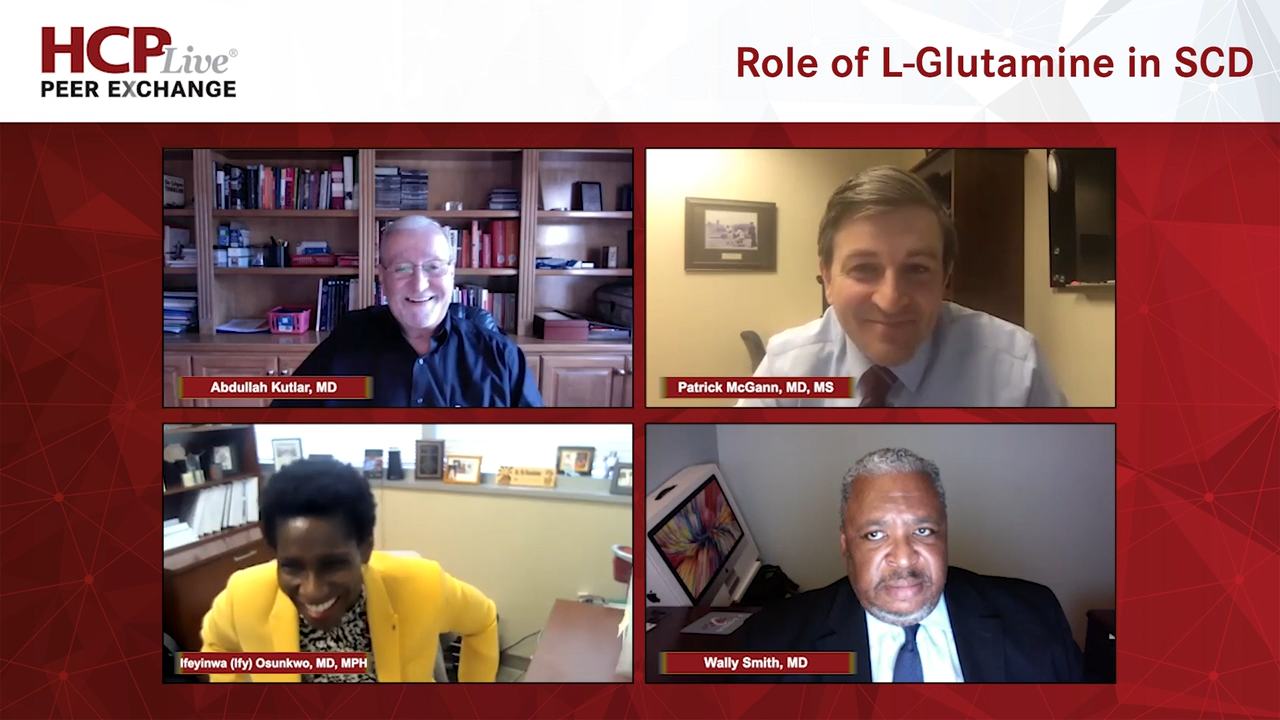


Ifeyinwa Osunkwo, MD, MPH: Hello, everybody, and welcome to HCPLive® Peer Exchange titled “Extra Perspectives on the Optimal Management of Sickle Cell Disease.” My name is Dr Ify Osunkwo, and I’m a hematologist and a professor of medicine in pediatrics at Atrium Health in Charlotte, North Carolina. I take care of a sickle cell [SC] disease population of about 1400 living with sickle cell disease in ages 18 and older. Today I have a few of my peers, and I would love to have them introduce themselves, starting with Dr Abdullah Kutlar.
Abdullah Kutlar, MD: Thank you, Ify. I’m Abdullah Kutlar, a hematologist and the director of the Sickle Cell Center at the Medical College of Georgia at Augusta University. I’m looking forward to this exchange.
Ifeyinwa Osunkwo, MD, MPH: Welcome, Abdullah. Wally?
Wally Smith, MD: Hi, I’m Wally Smith. I’m a general internist by training, but I’ve been taking care of patients with sickle cell disease since 1984 at 2 institutions. Since 1991, I’ve been at Virginia Commonwealth University, where I direct the Adult Sickle Cell Center.
Ifeyinwa Osunkwo, MD, MPH: Our next panelist is Dr Pat McGann. Pat?
Patrick McGann, MD, MS: I’m Pat McGann. I’m an associate professor of pediatrics and a sickle cell doctor at Cincinnati Children’s Hospital. I take care of many children with sickle cell disease in the United States and have lots of partnerships across the world, so I’m helping to improve outcomes globally as well.
Ifeyinwa Osunkwo, MD, MPH: Welcome, everybody. Our discussion is going to focus on providing an overview of sickle cell disease and discussing available standards of care using treatments of sickle cell disease. We’re also going to take a deep dive into the new treatments that were actually approved over the past 4 or 5 years, and our long-standing treatment for sickle cell disease that we’ve had for the past 20 years. Welcome, everybody. Let’s get started. Our first question is going to Wally. How do you define sickle cell disease to the medical audience and even the patient audience?
Wally Smith, MD: First, you must understand what hemoglobin is. Hemoglobin is a molecule inside your red blood cells that carries oxygen. You need hemoglobin to pick up the oxygen and deliver it to the tissues. Patients with sickle cell disease have a problem with that because they have a deficient hemoglobin. The hemoglobin doesn’t pick up oxygen well and doesn’t hold on to it well, and when it doesn’t hold on to oxygen, it tends to form funny red blood cells, like a garden sickle. The hemoglobin inside forms little crystals or polymers and stretches the cell from what normally is a doughnut into funny shapes, including the garden sickle. When you look at somebody’s blood who has sickle cell disease, you don’t see a bunch of doughnuts, you see these funny shaped cells.
There are 4 genetic kinds of sickle cell. It’s a genetic disease; mom and pop have to have the trait in order for Junior to get the disease. It’s not an every-time thing. Every time mom and pop have a child, there’s a 1 in 4 chance for Junior to get the disease. Some families are unlucky, 3 or 4 children in the family will have the disease because every time there’s a 1 in 4 chance.
There are 4 genotypes prevalent in the United States. Some would say 5, some would say 6 if you go worldwide. The more severe genotypes are SS [sickle cell anemia]. That’s where mom and pop have the same hemoglobin S [sickle] gene. And then S beta plus thalassemia, where mom or dad has the hemoglobin S gene and dad has the thalassemia gene that passes on. If you draw all those things out, the complications get significant, but the main thing to take away is it’s genetic and there’s a bad hemoglobin. People with this disease do not do well. They die early, they’re anemic, they’re in pain, they have infections, and they have strokes. It’s a miserable life, and until recently it was a childhood killer. Only recently have we been able to stretch the length of life past the childhood stage.
Ifeyinwa Osunkwo, MD, MPH: That is interesting, Dr Smith. Abdullah, can you highlight the differences in the genotypes? Many patients say they have a touch of sickle cell or the milder form. Can you explain to us what it means to have the different types of sickle cell disease that Wally had mentioned? There are 4 to 5 common types. Can you talk about that?
Abdullah Kutlar, MD: Wally made a great introduction and made it easy by mentioning the different genotypes of sickle cell disease. If you have 2 copies of the hemoglobin S gene, 1 from each parent, you have with hemoglobin S or sickle cell anemia. Then there are other mutations or variant hemoglobins, such as beta-thalassemia and hemoglobin C, which can interact with sickle hemoglobin to generate a phenotype of sickle cell disease.
If you look at the US population, I can cite some of the statistics from our adult population. This is about the same, give or take a few percentage points. About 69% of our patient population is hemoglobin S, followed by SC disease in about 20%, and S beta plus thalassemia in 6%. S beta zero thalassemia is in 3% of our patient population. There’s a 1% to 2% of those rarer genotypes, such as SD Los Angeles, SE, SOR1. The interesting thing is that for all practical purposes, the phenotypes of SS and S beta zero are similar, and they run a similar clinical course, as do SC and S beta plus thalassemia.
The Cooperative Study of Sickle Cell Disease [CSSCD], which is the largest natural history study conducted in upward of 20 centers in the United States between the late 1970s and early to mid-1990s, shows that if you look at the clinical features, SS and S beta zero cluster together, whereas SC and S beta plus cluster together. This was a 1994 publication in the New England Journal of Medicine by Ora Platt and other investigators from the CSSCD. The crisis or VOC [vaso-occlusive crisis] rate is twice in the SS, S beta zero group compared with SCS beta plus. The same applies for an acute chest, and retinopathy seems to be more common in SC, which is a viscosity-related pathology, so vascular necrosis. There are distinct differences, although these can be grouped into SS, S beta zero vs S beta plus, and SC.
There can still be some mild differences among these genotypes. It’s safe to say that SS and S beta zero are more severe phenotypes of the disease than SC and S beta plus.
Transcript Edited for Clarity


