Management of Beta Thalassemia - Episode 2
Prevalence and Clinical Presentation of Beta Thalassemia


Peter L. Salgo, MD: Tell me about the prevalence and the burden of this disease? How bad is it? Who’s got it?
Sujit Sheth, MD: So β-thalassemia is prevalent worldwide. It’s estimated that about 80 to 90 million people are carriers for one of these genes. That’s about 1% to 1.5% of the population.
Peter L. Salgo, MD: Stop for a minute. 1.5%. This is not a small problem; it’s a biggie.
Sujit Sheth, MD: These are carriers though.
Peter L. Salgo, MD: That’s okay; that gene is out there.
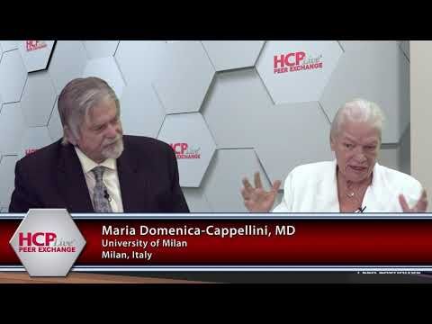
Sujit Sheth, MD: It’s out there and it’s estimated that in Italy, for instance, there are about 6000 affected individuals who have thalassemia, the disease. In the US [United States] it’s about 2000 or so. That’s an estimate. But there’s probably the major burden of it is in Asia.
Peter L. Salgo, MD: Really?
Sujit Sheth, MD: Which you find in the southeast Asian countries, the Indian subcontinent, and then all the way extending through China as well.
Peter L. Salgo, MD: That’s interesting, because when I was in medical school—pterodactyls flew through the sky—it was always presented as a Mediterranean problem.
Maria Domenica-Cappellini, MD: Yes.
Peter L. Salgo, MD: Italians had β-thalassemia more than others.
Maria Domenica-Cappellini, MD: It is Italian based, but do you know why?
Peter L. Salgo, MD: Why?
Maria Domenica-Cappellini, MD: That is the distribution: malaria.
Peter L. Salgo, MD: Is that why?
Maria Domenica-Cappellini, MD: Because malaria was a selective natural factor, and the carriers of the β-thalassemia gene were surviving through malaria. So if you overlap the map of malaria in the world and the map of β-thalassemia distribution, they are exactly overlapping.
Peter L. Salgo, MD: It’s the same as sickle cell anemia then, right?
Maria Domenica-Cappellini, MD: Exactly.
Peter L. Salgo, MD: These are survival adaptations, which, unfortunately, have some downsides.
Maria Domenica-Cappellini, MD: That’s also true for other red blood cell defects. Because the defective red blood cells survive.
Peter L. Salgo, MD: So how does this disease present? How do you work it up? How do you recognize it in your office or in your clinic?
Thomas D. Coates, MD: Well, I think the presentation probably depends a lot on what part of the world…you’re in. I mean, classically, these children present at several months of age. You know initially when they’re born they have fetal hemoglobin. And then they transfer.
Peter L. Salgo, MD: That’s protective.
Thomas D. Coates, MD: Protective. Because the fetal hemoglobin is α chains and γ chains, and then you switch to β chains and β-thalassemia somewhere around 2 to 3 months of age and the patient begins to become anemic.
Peter L. Salgo, MD: It’s not an accident that it’s β-thalassemia. β chain is where the problem is.
Thomas D. Coates, MD: Yeah, right. I mean there is a γ-thalassemia, but it’s pretty rare. So they present with anemia. Now there’s over 300 mutations in the β chain that can cause this, and so it’s a very broad range of severity of how these kids present. But, generally, they present with anemia in early infancy.
Peter L. Salgo, MD: What about growth failure?
Farzana Sayani, MD: Well, again, it depends a lot upon what kind and when they become anemic and how anemic they are. And so normally when they present to the physician, it’s because of anemia. They could have big spleens because the body is trying to make more red blood cells, make more hemoglobin, but it can’t. So it reverts to making hemoglobin in the spleen and other places outside of the bone marrow.
But anemia is primarily the presentation. Now whether they have growth failure or not depends again on the severity. If the patient’s hemoglobin level isn’t so bad, the growth may not be so bad. I mean, normal hemoglobin is around 12, let’s say. So if the baby’s hemoglobin is at 9 or 10, they won’t be so affected. If they [have] thalassemia major and their hemoglobin is 5, then they’ll begin to have growth failure right away.
Peter L. Salgo, MD: What about effects on the heart? What about thrombotic problems with it?
Farzana Sayani, MD: Well, again, when you’re talking about presentations, these things occur when the patients are much older and these classical things that people think of, it depends. And again, maybe that’s a problem with the 4 of us here is we know what too many of these depending kinds of things are.
Certainly when the patients are older they have problems with thrombosis and they have a lot of other difficulties. But usually we interfere, intervene with treatment. Now a lot of places in the world, especially in the Western world, the clinical presentation is at birth because people have newborn screening programs. And so, in the United States, for example, and in California, all the babies are screened. And so we get a letter from the state of California within days or weeks of birth notifying the physician, notifying the parents, notifying the local β-thalassemia centers that the child was born and they recommended that these kids then go in to be seen. So they’re diagnosed really even before they develop symptoms.
Peter L. Salgo, MD: But I take from what you’re saying that when you talk about cardiac abnormalities, you’re talking about thrombosis, these are later. And early on what you’re really looking at is the anemia, the hemoglobin.
Thomas D. Coates, MD: So before people learned to start transfusing these patients, you know, the children usually didn’t survive…the severe ones. Again, this is very important, remember there are 300 different genes just for purpose of the discussion, which means there are 300 from the mother and 300 from the father, so there’s a lot possibility.
Peter L. Salgo, MD: It’s nominal.
Thomas D. Coates, MD: A lot of possible combination here. But in the kids that had hemoglobin in the 4 or 5 range, I mean, they usually didn’t make it to 5 or 6 years of age. Then people figured out that if you routinely transfused the patient, they would survive much longer. But then, because transfusion gives you iron, they developed iron overload and the median survival in the ‘60s and ‘70s was around 15 years of age, approximately. And there they would die from heart failure, or mainly from heart failure: the iron overload. Well it’s primarily the cardiac, the liver certainly is involved, but the big problem was iron cardiomyopathy and, of course, endocrine failure.
Transcript edited for clarity.